MRI scans provide detailed images of soft tissues and organs, making them invaluable in cancer diagnosis, staging, and treatment planning. Learn how MRI helps detect tumors and monitor therapy response.
What is an MRI Scan?
MRI stands for Magnetic Resonance Imaging. It uses powerful magnets and radio waves to create highly detailed cross-sectional images of the inside of the body, particularly soft tissues like the brain, spinal cord, muscles, and internal organs.
Unlike CT scans or X-rays, MRI does not use ionizing radiation, making it safer for repeated use.
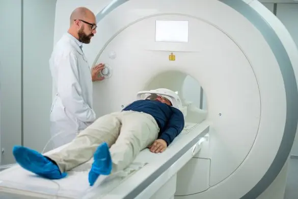
Is an MRI Painful?
No, an MRI scan itself is not painful. However, lying still inside the narrow tube for 30–60 minutes can be uncomfortable for some, especially those with claustrophobia. Open MRI machines are available for such patients.
You may hear loud knocking sounds during the scan; earplugs or headphones are usually provided.
Is a CT Scan the Same as an MRI Scan?
No. While both produce detailed body images, they work differently:
- CT Scan: Uses X-rays to generate images. Faster than MRI, better for visualizing bones and acute bleeding.
- MRI: Uses magnetic fields and radio waves. Superior for soft tissue contrast, ideal for brain, spine, pelvic organs, and ligaments.
In some types of cancer, like brain, spinal cord, or cervix, an MRI is preferred over a CT scan because it can produce clearer results.
How Does an MRI Scan Help a Cancer Patient?
Your oncologist might use an MRI scan for one or more of the following reasons:
- To find the location of a tumour in your body
- To determine the size of the tumour and whether it has spread (helps in staging cancer)
- To assess if a tumour is cancerous or benign
- To check whether treatment is working (by comparing pre- and post-treatment scans)
- To guide biopsies or surgery
Based on this information, your oncologist can plan the next steps of your cancer treatment effectively.
Preparing for an MRI Scan
- Inform your doctor if you have any metal implants (pacemakers, cochlear implants, aneurysm clips), as they may interfere with the machine.
- You may be asked to remove jewelry, hearing aids, dentures, or clothing with zippers/metal parts.
- In some cases, a contrast agent (gadolinium-based) is injected intravenously to enhance image clarity.
- Fasting is usually not required unless specified.
Specialized MRI Techniques in Oncology
- Functional MRI (fMRI): Maps brain activity before neurosurgery.
- Diffusion-Weighted Imaging (DWI): Detects changes in water movement within tissues, useful in identifying aggressive tumors.
- MR Spectroscopy: Analyzes chemical composition of tissues to differentiate tumor types.
- Dynamic Contrast-Enhanced MRI: Monitors blood flow patterns in tumors.
Advantages and Limitations
Advantages:
- No radiation exposure
- Excellent soft tissue resolution
- Multiplanar imaging capability
Limitations:
- Longer scan time
- Not suitable for patients with certain metallic implants
- Higher cost compared to CT
Follow-Up Scans
Repeat MRIs are often used during and after treatment to monitor tumor response, detect recurrence, or evaluate new symptoms. Consistent imaging protocols allow accurate comparison over time.