Understanding the different types of mammograms—2D, 3D, contrast-enhanced, and digital breast tomosynthesis—is key to early breast cancer detection. Learn how each works and their benefits.
Here’s a look at the most common types of breast cancer imaging screening tests available today. Apart from these, breast cancer self-examination (self-checking for lumps) and clinical breast examination (breast examination by treating physicians) are also important in catching cancer in the early stages, while it is still easily treatable.
The goal of breast cancer screening is to find cancer early, before it has a chance to grow, spread, or cause problems.
What is Mammography?
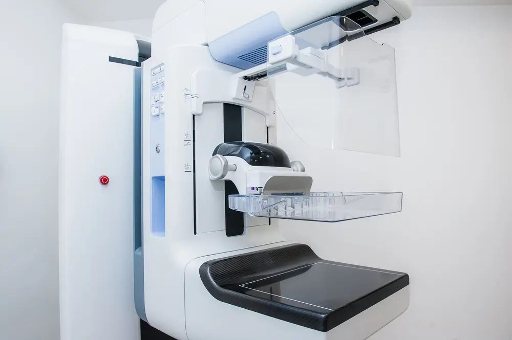
Mammography is an imaging technique that uses low-dose X-rays to examine the human breast. It's a critical tool for early detection of breast cancer, often identifying tumors before they can be felt during a physical exam.
Types of Mammography
2D Mammography (Digital Mammography)
This traditional form captures two X-ray images of each breast from different angles—top-to-bottom and side-to-side. While effective, overlapping tissue can sometimes make it harder to detect abnormalities, especially in women with dense breasts.
3D Mammography (Digital Breast Tomosynthesis)
Also known as tomosynthesis, this advanced technique takes multiple X-ray images from various angles around the breast. A computer then reconstructs them into a 3D image, allowing radiologists to view breast tissue layer by layer.
Benefits:
- Improved accuracy in detecting cancers
- Reduced false positives and fewer recall rates
- Better visualization in women with dense breast tissue
Contrast-Enhanced Spectral Mammography (CESM)
This newer type combines a standard mammogram with a contrast agent (iodine-based dye) injected intravenously. The contrast highlights areas of increased blood flow, which are often associated with cancerous growths.
Best for: Evaluating suspicious findings, monitoring treatment response, and assessing high-risk patients.
Screening vs Diagnostic Mammography
Screening Mammography: Performed routinely on women without symptoms to detect early signs of cancer.
Diagnostic Mammography: Used when a woman has symptoms such as a lump, pain, nipple discharge, or after an abnormal screening result. Provides more detailed images.
Who Should Get Screened?
Guidelines vary slightly by country and organization, but general recommendations include:
- Women aged 40–74 should undergo regular screening mammograms.
- Those with a family history or genetic predisposition (e.g., BRCA mutations) may start earlier and require additional imaging like MRI.
How to Prepare for a Mammogram
- Avoid using deodorants, powders, or lotions on the day of the test, as they can interfere with the images.
- Wear a two-piece outfit for easier undressing.
- Schedule the test one week after your period when breasts are less tender.
- Bring prior mammogram results if changing facilities.
Limitations and Considerations
While mammography is highly effective, no test is perfect:
- Dense breast tissue can mask tumors.
- False positives can lead to unnecessary anxiety and biopsies.
- Overdiagnosis and overtreatment of slow-growing cancers remain concerns.
In some cases, supplemental screening with ultrasound or MRI may be recommended based on individual risk factors.
Why Early Detection Matters
Not all pains or lumps in the breast are indicative of cancer. Even if there is a lump, it may not always be cancerous. But there is no way to know except to visit a doctor and get a proper diagnosis.
The more you delay, the more the cancer grows and moves into advanced stages. When cancer spreads to other organs, it becomes more challenging to treat successfully.
Visit your doctor at the earliest and speak about getting a mammography done.